
Research
Psychiatry still relies largely on what patients report about their symptoms to diagnose and treat mental disorders. But two people with the same diagnosis can look very different, while the same symptoms can occur across different disorders, making misdiagnosis possible. Treatment decisions also largely follow general guidelines rather than information measured in the individual patient. This can mean delays in finding the right treatment, or receiving a treatment that was never likely to work.
My research asks whether measurements of the brain can add something useful.
Can we sharpen diagnosis, explain why some people develop psychiatric disorders while others do not, and predict which treatment will work for a given patient?
I work across obsessive-compulsive, anxiety and mood disorders, using brain scans from thousands of people, machine learning, and studies that follow the same individuals over many years.
Four strands run through this work.
Brain development and individual trajectories
Many psychiatric disorders first appear during childhood, adolescence or young adulthood, while the brain is still changing rapidly. Yet much of psychiatric brain imaging relies on single time-point measurements and group averages: patients are scanned once and compared with healthy people, rather than tracking how an individual brain develops over time. This tells us how groups differ, but not how an individual got there or where they may be heading.
Normative modelling offers a way to shift the focus from group averages to the individual. Like growth charts used in child health, it maps the range of brain measures expected at each age and asks where an individual sits within that range. The degree to which someone deviates from what is expected can then be measured directly.
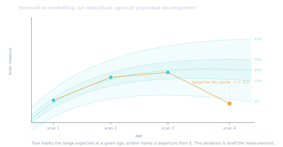
With repeated scans of the same people, this approach can do something a snapshot cannot: follow individuals along their own developmental paths, and ask whether deviations in brain development come before symptoms appear, change alongside them, or follow them. That ordering in time is essential for understanding what drives what.
This is the focus of my current work at Amsterdam UMC with Prof. dr. Odile van den Heuvel, using Generation R, a Dutch study that has followed thousands of children from before birth with repeated MRI scans. Two preregistered projects run in parallel: one examining the maturation of the thalamus, a relay station deep in the brain, in relation to wider brain development, and another examining brain maturation in relation to obsessive-compulsive symptoms.
At Leiden University, I work with Dr. Moji Aghajani on similar questions in adolescent anxiety, using data from the ENIGMA-Anxiety consortium.
Related work uses brain-age modelling. A model learns to guess a person’s age from their brain scan alone; applied to a new individual, the gap between the guessed age and their real age becomes a simple summary of whether their brain looks older or younger than expected.
A KNAW Ter Meulen Beurs supports an international extension of this line of research with the University of Southern California and the University of Michigan. Using scans from roughly 15,000 young people across more than thirty countries, this work aims to develop reference curves that capture normative variation in brain development across the population, and identify individual deviations across the full spectrum of mental health disorders.
Diagnostic biomarkers in OCD and anxiety
Can a brain scan tell us whether someone has a psychiatric disorder?
Working with the ENIGMA-OCD consortium, I tested this on the largest dataset then available: scans of brain anatomy from 2,304 people with OCD and 2,068 healthy controls, collected at 36 institutes around the world.
The result was informative in an unexpected way. Models trained to recognise OCD from brain anatomy did not hold up when tested on scans from hospitals they had not seen before. But models distinguishing medicated from unmedicated patients worked considerably better, revealing widespread differences in brain anatomy associated with medication use. The lesson: the differences between patients, including the treatments they are already receiving, can be larger than the differences produced by the disorder itself.
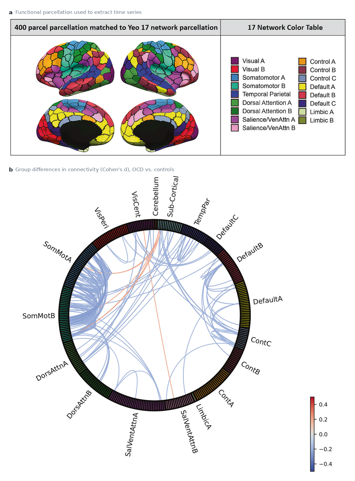
A follow-up study looked at brain activity rather than anatomy, measuring how strongly different regions communicate with one another in more than 2,000 participants. People with OCD showed weaker communication across much of the brain, and the effect was strongest in the sensorimotor network, the regions handling movement and bodily sensation, rather than in the decision-and-habit circuits that textbook models of OCD emphasise. The disorder’s biology, in other words, may reach further than the standard models suggest.
I then extended this approach to anxiety disorders in young people, using scans from 3,343 participants aged 10 to 25 across 32 sites. Models that combined information from across the whole brain could separate patients from controls only modestly, though far better than any single brain measure could. Together these studies show both the promise of brain-based classification and how far it still is from being clinically useful.
Key papers
-
Han, Bruin, et al. (2025). Structural brain differences associated with panic disorder: an ENIGMA-Anxiety Working Group mega-analysis of 4,924 individuals worldwide. Molecular Psychiatry. Shared first author. Link
-
Bruin et al. (2024). Brain-based classification of youth with anxiety disorders: transdiagnostic examinations within the ENIGMA-Anxiety database using machine learning. Nature Mental Health. doi:10.1038/s44220-023-00173-2
-
Bruin et al. (2023). The functional connectome in obsessive-compulsive disorder. Molecular Psychiatry. doi:10.1038/s41380-023-02077-0
-
Bruin et al. (2020). Structural neuroimaging biomarkers for obsessive-compulsive disorder in the ENIGMA-OCD consortium: medication matters. Translational Psychiatry. doi:10.1038/s41398-020-01013-y
Predicting treatment outcome
Diagnosis is only one challenge. Clinicians usually know who is unwell. What they cannot know in advance is which treatment is likely to work for which patient. Choosing the wrong treatment can cost months, years, or, in some cases, a life.
My clearest example is electroconvulsive therapy (ECT), one of the most effective treatments for patients with treatment-resistant depression (TRD), who have not responded to prior medication and therapy. Working within the Global ECT-MRI Collaboration (GEMRIC), I built models that combine brain structure, brain activity and clinical information to predict, before treatment begins, who will recover. At the larger contributing centres these predictions were right substantially more often than chance, and crucially they remained informative when tested on hospitals the models had never seen.
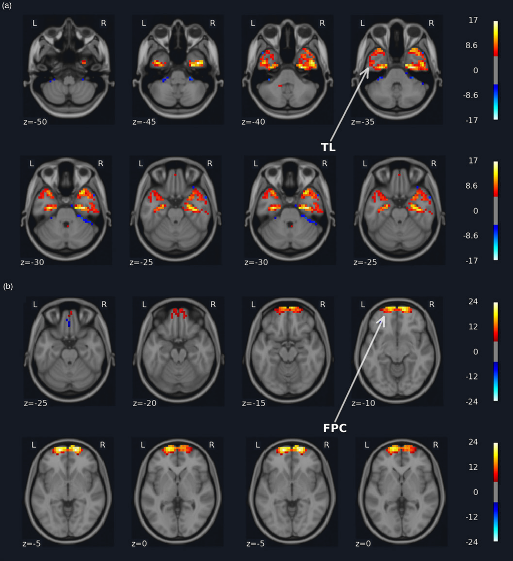
ECT is a setting where a prediction like this could genuinely matter: the treatment is demanding for patients and hospitals alike, its effects when it works are substantial, and an MRI scan is cheap compared with the cost and burden of a full treatment course. It is a concrete example of where a validated brain measure could one day help decide who should be offered which treatment.
The same question applies to psychological treatment. In related work I contributed to models predicting cognitive behavioural therapy outcome in OCD from clinical and neuroimaging data, where clinical variables turned out to carry much of the predictive information. That result is a useful corrective: neuroimaging has to earn its place against simpler and cheaper measures rather than being assumed to improve on them.
I have also conducted a randomised controlled trial in OCD examining how pharmacological and psychological treatment affect brain activity, from recruitment through data collection.
Most recently, I am a co-investigator on a project funded by the ZonMw Neuropsychoanalyse Fonds investigating the brain dynamics underlying esketamine treatment in depression. Esketamine acts far more rapidly than conventional antidepressants, which makes it an unusually informative setting for asking what changes in the brain when a treatment works, and how quickly.
Key paper
- Bruin et al. (2023). Development and validation of a multimodal neuroimaging biomarker for electroconvulsive therapy outcome in depression: a multicenter machine learning analysis. Psychological Medicine. doi:10.1017/S0033291723002040
Making biomarkers generalisable
It is surprisingly easy to build a model that predicts well for the patients it was developed on and then fails for patients at a different hospital, scanned on a different machine. This is one of the central challenges in psychiatric neuroimaging, and much of my methodological work confronts it directly.
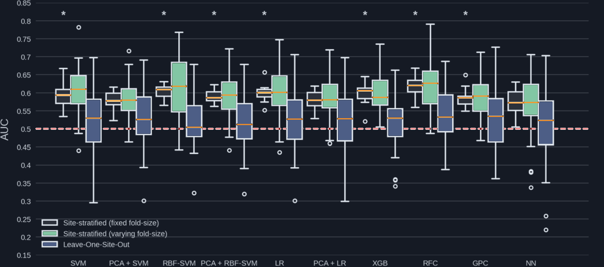
Data pooled from dozens of hospitals arrive with dozens of differences: different scanners, different scanning protocols, different patient populations, different diagnostic habits. Rather than treating all of that variation as a nuisance, my work examines how it shapes the measurements and predictions we make, and what it takes for a finding to hold up in spite of it.
For ENIGMA-OCD I developed the consortium’s first shared framework for analysing brain activity data across all of its sites at once; it has since been adopted by more than ten other ENIGMA working groups studying different disorders. I have also worked on the quieter problems that decide whether such analyses can be trusted: missing data, scanner differences between sites, and how models should be validated.
The principle running through all of it is external validation: testing every model on data it has never seen, from places it has never been, and reporting openly when it fails. That is a far stronger test than any amount of checking a model against the data it was built from.
My analysis code and preprocessing pipelines are openly available where possible. Making methods inspectable and reproducible is part of the same goal: developing neuroimaging biomarkers that are not only statistically interesting, but robust enough to be useful beyond the dataset in which they were developed.
Towards individualised psychiatry
What connects these projects is a focus on understanding psychiatric illness at the level of the individual.
Rather than asking only whether a disorder is associated with an average difference in the brain, I want to understand how brain development differs between people, how these differences relate to symptoms, and whether they can provide information about prognosis or treatment response.
Achieving this will require following people over time rather than photographing them once, measuring more than one aspect of brain and behaviour, working with large and diverse samples, and validating every model far beyond the data it was built on. The goal is not prediction for its own sake, but brain measures that are reproducible, generalisable and clinically meaningful.
A full and continuously updated list of my work is available on the publications page. More about my background is on the about page.